|

|
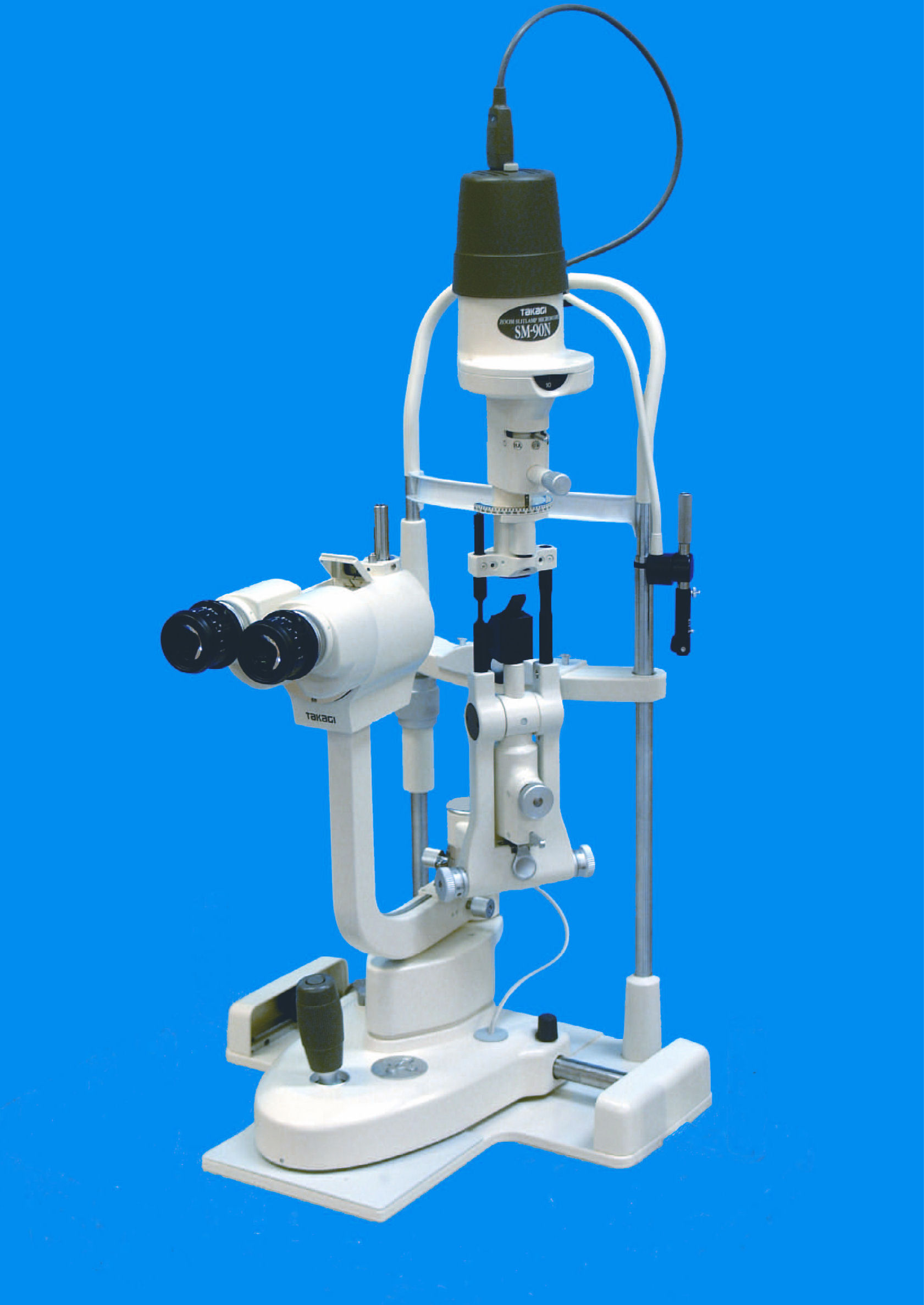
SM-90N
Zoom Slitlamp
Microscope |
|
|
 |
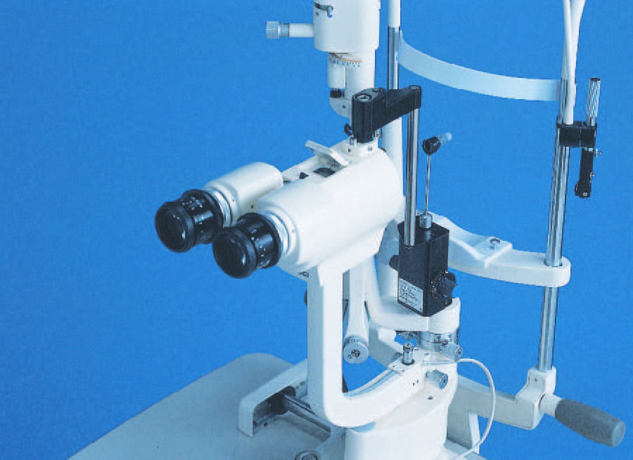
High-resolution microscope with
motorized zoom system
The TAKAGI
slitlamp technology is combined
with an electric zoom mechanism
unique in its class. The zoom
mechanism allows magnification
to be changed over a range of
5.5x to 32x to provide optimum
magnification in clinical
applications. All lenses
employed in the microscope are
high-quality multi-coated for
clear and bright images. |
|
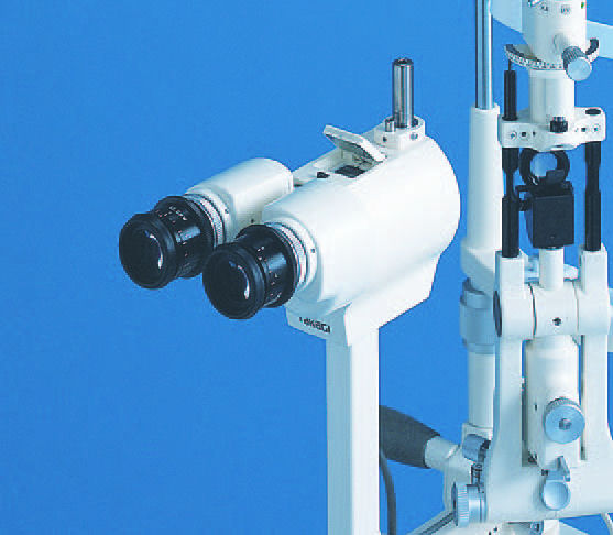
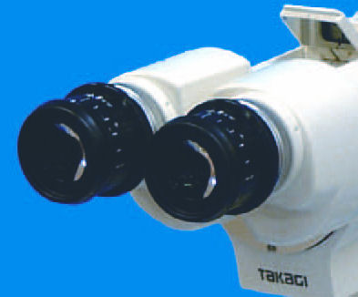
Eyepiece with helicoid mechanism
for diopter adjustment
The 12.5x high-eyepoint
eyepieces with an expanded field
of view enable observation over
a wider area. With the diopter
adjustment system that employs a
helicoid mechanism, the diopter
can be adjusted without rotating
the lenses or the eye cap. This
feature has been well received
as it prevents the adjusted
diopter from accidentally being
changed during use as the eye
caps will not rotate. |
 |
 |
Magnification display using
one-touch flip-up mirror
The current
magnification is displayed using
the one-touch flip-up mirror,
thus allowing photography at the
fixed magnification when taking
multiple images. Interior
illumination of the display unit
ensure that the magnification
display is visible even in dark
surrounding. Fitting the new
combination adapter (S10-17)
ensures that the magnification
is displayed even when using an
imaging system. |
|
Special mirror coating and
diffuser The
mirror applying a special
coating eliminates almost all
ultraviolet and infared lights
to improve protection against
phototoxicity for the examinee's
retina. At the same time,
natural images are obtained in
the visible light spectrum,
being improved in comparison
with the UV filter (TAKAGI
comparison). The use of the
standard diffuser allows
illumination over a wider range
when photographing
the anterior segment of the eye. |
 |
 |
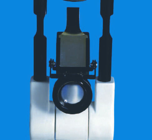
Tonometer mount
The tonometer
mount is fitted to the top of
the microscope as standard, and
fitting the TAKAGI applanation
tonometer (AT-1) allows
measurement of intraocular
pressure. |
|
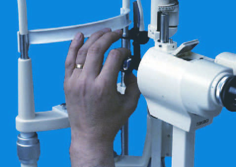
New form headrest
The new form
headrest functions not as a
headrest for the examinee but
also as a support for the
examiner holding an indirect
lens upon fundus examination,
reducing the fatigue in the arm
caused by long hours of
examination. |
 |
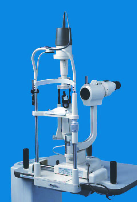 |
Slitlamp with integrated base
By integrating it
with the base, the sturdiness of
the chin rest assembly has
improved dramatically. Now that
the base is integrated, there is
no need to be selective with the
sharp of fittings for the chin
rest assembly or its
installation method. The
slitlamp can now be set up very
easily on any type of instrument
table.
|
| |
Right eye / Left eye recognition
sensor and signal output
faunction The
right eye/left eye recognition
sensor is now buit-in so that
the slitlamp works well with an
image filing system. Right
eye/left eye recognition signal
is output once the slitlamp is
aligned to the eye to be tested.
* The
cable-end connector of the
connecting cable (optional)
varies according to the image
filing system used. |
| |
Centralized control system
In addition to
the ability to move the slitlamp,
and 3D movement in the X,Y, and
Z directions by joystick the
provision of a trigger button
(also functions as the light
boost button) at the top, and
connection to video equipment
allows the examiner to acquire
excellent images while looking
through the slitlamp. The newly
developed X-Y control button
fitted for the first time to the
slitlamp allows the zoom up-down
and the intensity increase and
decrease to be controlled with
one hand. The X-Y control button
may be rotated 90 degrees, thus
allowing the examiner to change
the direction as necessary.
* Light boost
and trigger functions do not
work simultaneously |
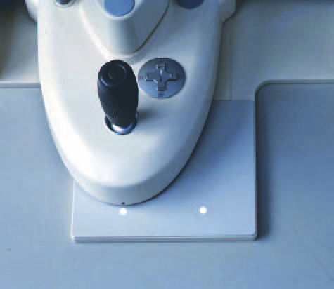 |
Navigation LED's
The LED's
illuminate to indicate the
approximate position to assist
focusing on the eye to be
tested. By aligning the marker
located on the base of the
slitlamp to the position of the
relevant LED, the microscope can
easily be focused on the right
or left eye.
* The focal
length between the microscope
and the eye to be tested varies
from individual to individual.
This function only provides
approximate positioning. |
Motorized zoom Slit lamp
Microscope.
|
MICROSCOPE |
|
CROSS-SLIDE BASE |
|
|
Type |
Galilean-type coverging
binocular microscope |
Longitudinal (coarse) movement |
90mm |
|
Magnification Changer |
motorized zoom |
lateral (coarse) movement |
110mm |
|
Eyepieces |
12.5x wide |
Horizontal (fine) movement |
15mm |
|
Total magnifications |
5.5x to 32x |
Vertical movement |
±15mm |
|
Real field of view |
40.9mm to 6.8mm dia. |
CHIN REST |
|
|
Interpupilary adjustment |
52mm to 85mm |
Vertical movement |
70mm |
|
Diopter adjustment tange |
-5 dioptor to +5 dioptor |
Fixation light |
LED (red) |
|
|
|
|
|
|
Slit width |
0 to 10mm,
continuously variable (at
10mm,slit becomes a circle) |
Input voltage |
100VAC to 230VAC
50/60Hz |
|
Slit length |
1 to 10mm,
continuously variable |
Maximum power
consumption |
64VA |
|
|
Aperture diaphragms |
10mm, 5mm, 3mm, 2mm, 1mm,
0.2mm dia. |
DIMENSIONS &
WEIGHT |
|
diaphragms Filters |
Heat absorbing, UV,
red-free, and cobalt blue |
Base dimensions |
359mm(W) x364mm(D) |
|
Lamp |
12V 30W halogen bulb |
Weight |
13.5kg |
|
|